Exercise Erased More Than Half the Molecular Signature of Muscle Aging. A New Study Maps Exactly Which Half.
Prefer to listen? Hit play for a conversational, audio‑style summary of this article’s key points.
A new study solved a problem that has undermined most exercise-and-aging research: separating aging from inactivity. Older people move less, so it is usually impossible to tell whether aging muscle reflects age itself or decades of reduced movement. This Nature Aging study from Amsterdam UMC and Maastricht University recruited older adults whose daily step counts and high-intensity activity matched young adults, isolating aging from inactivity, then compared them to older adults who had trained consistently for years and to older adults with early physical impairment.
The defining molecular signature of muscle aging is an energy crisis. Comparing young adults to activity-matched older adults, 1,106 genes were downregulated with age, dominated by genes for cellular respiration and energy metabolism, including the ATP synthase, cytochrome c oxidase, and NADH dehydrogenase subunits that build the mitochondrial energy machinery. This was accompanied by declining NAD+ and accumulating triglycerides inside the muscle. The tissue was losing its ability to make energy while storing more unburned fuel.
More than half of the molecular signature of muscle aging was absent in trained older adults. Specifically, 55.9 percent of age-related upregulation and 57.1 percent of downregulation were missing in the trained group, with sensitivity analyses putting the range at 45 to 62 percent. Their muscle resembled that of young adults far more than their chronological age would predict.
The changes training preserved were specifically the energy metabolism ones. The single most prominent feature of muscle aging, the decline in cellular respiration genes, turned out to be the single most preventable. Genes like NDUFS1 and COX5A were depleted in normally active and impaired older adults but sat at youthful levels in the trained, across all five mitochondrial respiratory complexes.
Being generally active was not enough. Structured training was the difference. The normally active older adults were walking as much as the young adults, and their energy metabolism genes declined anyway. What preserved the youthful profile was structured, sustained training, at least three hour-long sessions per week for over a year. This distinction matters: filling a step counter and being genuinely trained are not equivalent at the molecular level.
Roughly half of muscle aging persisted regardless of training, and this "unavoidable" half is where drugs will have to work. Changes in synaptic transmission genes like PCDH8 and UNC13C, and in WNT signaling genes like DAAM2 and CTR9, appeared in all older adults. The authors frame this as the division geroscience should organize around: lifestyle for the preventable half, therapeutics for the unavoidable half. Notably, the unavoidable changes did not cluster into a single clear target, so the work of finding one is still ahead.
The fittest muscle mounted the largest inflammatory response to exercise, and that response tracked with better health. All groups activated stress and immune genes after exercise, including IL6, IL1B, and TNF, but the magnitude scaled with fitness. Trained older adults most closely resembled young adults in their response (r = 0.451), followed by normally active (r = 0.394), with impaired older adults the most blunted (r = 0.263). The stress response to exercise appears to be the mechanism of adaptation, not damage to be minimized.
This raises a genuine concern about anti-inflammatory longevity strategies. If the inflammatory stress response is how exercise produces its benefits, chronically suppressing inflammation, whether through IL-11 inhibitors or routine anti-inflammatory agents, may blunt the adaptation that exercise depends on. The authors raise this directly. It does not mean inflammation is beneficial in general, but it suggests the timing and context matter, and that suppressing the acute exercise response may carry a hidden cost.
A separate discovery identified the proteasome as a previously unrecognized regulator of NAD+. Genes negatively correlated with NAD+ were enriched for protein degradation pathways. Testing this directly, the proteasome inhibitor MG-132 raised NAD+ in both muscle and liver cells to a degree comparable to the precursor supplement NMN. This does not make proteasome inhibition a viable strategy, since the proteasome is essential, but it opens a new route to understanding NAD+ decline that operates through protein turnover rather than supplying more raw material.
Introduction
There is a problem that has quietly undermined most of what we think we know about exercise and aging. Older people move less. On average, they take fewer steps, spend less time at high intensity, and sit for longer stretches than younger people do. So when a study compares the muscle of a seventy-year-old to the muscle of a twenty-five-year-old and finds that the older muscle has fewer mitochondria, lower energy production capacity, and a deteriorated molecular profile, there is an obvious question that the study usually cannot answer: is that aging, or is that just twenty years of not moving very much?
The distinction matters enormously. If the molecular deterioration of aging muscle is largely the accumulated consequence of inactivity, then it is addressable, and the intervention is obvious. If it is aging itself, an intrinsic biological process that proceeds regardless of behavior, then no amount of exercise will touch it and the field needs to be looking for drugs. Most studies cannot separate these two possibilities because the confound is baked into their design.
A new study published in Nature Aging from a team at Amsterdam UMC and Maastricht University solved this problem with an unusually careful piece of cohort construction. They recruited young adults and older adults whose daily step counts and time spent in high-intensity activity were comparable to each other. Not older adults who were sedentary. Older adults who were moving just as much as the young controls. Any molecular difference between those two groups is therefore attributable to aging rather than to inactivity.
Then they added a third group: older adults who had been training seriously and consistently for years, defined as at least three structured hour-long exercise sessions per week for an uninterrupted period of more than a year. And a fourth: older adults with measurable physical impairment, the beginnings of frailty. They took muscle biopsies from all of them, before and immediately after a one-hour bout of cycling, and ran transcriptomics on more than 24,000 gene transcripts, metabolomics on 135 metabolites, and lipidomics on 1,383 lipid species.
What emerged is the closest thing the field has produced to a molecular answer to a question people have been asking for decades: how much of muscle aging is actually optional?
The answer, it turns out, is more than half. But not all of it. And the parts that training could not touch are as informative as the parts it erased.
The Cohort and Why Its Design Is the Whole Point
The strength of this study lies almost entirely in who was in it, so it is worth understanding the four groups before looking at what was found in them.
The young group consisted of 11 adults in their twenties, serving as the reference point for what muscle looks like before aging has acted on it. The older participants, 36 in total, were divided into three groups that together capture the full range of how muscle ages depending on how a person has lived.
The first older group, and the analytical anchor of the entire study, was the normally active group: 15 older adults whose daily physical activity matched the young controls. This matching is the design decision that makes everything else interpretable. Their step counts were comparable to the young group. Their time spent in higher-intensity activity was comparable. These were not sedentary older people. They were living physically ordinary lives at a level of daily movement equivalent to adults five decades younger. Which means that when their muscle is compared to the young group's muscle, whatever differences appear cannot be attributed to the older group simply moving less. The differences are aging itself, isolated from inactivity for perhaps the first time in a study of this molecular depth.
The second older group was the trained group: 16 older adults who had committed to structured exercise, at least three supervised hour-long sessions per week, sustained without interruption for more than a year, and in many cases far longer. This is the group that reveals what deliberate, consistent training on top of an already active baseline actually buys a person at the molecular level. The comparison that matters is not trained older adults against sedentary ones. It is trained older adults against older adults who were already moving as much as the young, which isolates the specific contribution of structured training from the contribution of general daily activity.
The third older group was the physically impaired group: 5 older adults with a Short Physical Performance Battery score of 9 or below, a validated threshold indicating meaningful decline in lower-body function and an elevated risk of progressing toward frailty and disability. This group is small, which is a genuine limitation we will return to, but it provides a window into what muscle looks like as it moves in the wrong direction, the molecular counterpart to the trained group's trajectory.
The comparisons this design enables are what give the study its power. Young versus normally active isolates aging from inactivity. Normally active versus trained isolates the effect of structured training from the effect of ordinary daily movement. And impaired versus everyone else shows what the decline toward frailty looks like beneath the surface. Each of these comparisons answers a different question, and the study was built specifically to be able to ask all three.
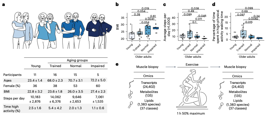
Figure 1: Cohort design and the energy metabolism signature of aging. Young and normally active older adults had comparable daily step counts and time in high-intensity activity, isolating aging from inactivity. Muscle biopsies were taken before and after a one-hour cycling bout and profiled by transcriptomics, metabolomics, and lipidomics. Among the genes downregulated with aging, those for cellular respiration, including ATP synthase, cytochrome c oxidase, and NADH dehydrogenase subunits, were the dominant signature.
What Aging Does to Muscle at the Molecular Level
Before we can understand what training preserved, we need to understand what aging took. And the first comparison in the study, young adults against the normally active older adults matched to them for daily movement, gives us the cleanest picture available of what aging does to muscle when inactivity is taken out of the equation.
The dominant signal was unambiguous. Among the genes that changed with age, 1,106 were downregulated, and the group of downregulated genes was heavily enriched for one biological theme above all others: cellular respiration and energy metabolism. These are the genes that build and operate the machinery of the mitochondria, the structures inside the cell that convert fuel into usable energy. When their expression falls, the muscle's fundamental capacity to produce energy falls with it.
To understand why this matters, it helps to understand what these genes actually do. The process of extracting energy from food and converting it into ATP, the molecule that powers essentially every active process in the cell, runs through a series of protein complexes embedded in the inner membrane of the mitochondria, collectively called the electron transport chain. The genes that were downregulated with aging included the subunits of these complexes. NADH dehydrogenase components, the NDUF genes, which form the entry point of the chain. Cytochrome c oxidase components, the COX genes, which sit near the end. ATP synthase subunits, the ATP genes, which perform the final step of actually manufacturing ATP. Aging was turning down the expression of the machinery at multiple points along the entire energy production pathway simultaneously.
This transcriptional finding did not stand alone. It aligned with what this same cohort had already been shown to exhibit in prior work: reduced mitochondrial mass, reduced mitochondrial respiration, and depletion of NAD+, a molecule central to energy metabolism that declines with age across many tissues. The metabolomic data in the current study confirmed the picture from a different angle, with NAD+ pathway metabolites appearing among the compounds most altered by age. The muscle was not simply expressing fewer energy genes. It was measurably losing the metabolic capacity those genes support.
The lipidomic data added a final dimension. Several triglyceride species were among the most age-altered lipids, all increased with age. Triglycerides are the storage form of fat, and their accumulation inside muscle tissue is a hallmark of metabolic aging. The muscle was becoming less able to burn fuel for energy and simultaneously accumulating more fuel in storage, a combination that captures the metabolic dysfunction of aging muscle in a single contrast: declining capacity to produce energy, rising deposits of unburned fat.
Put simply, the defining molecular signature of muscle aging in this study was an energy crisis. The genes for making energy were being turned down, the metabolic markers of energy production were falling, and fat was accumulating in the tissue that was losing its ability to burn it. This was the state of the normally active older adults, people who were walking as much as adults fifty years younger. It is the picture of aging with inactivity removed. The question the study was built to answer next was how much of this picture structured training could change.
The Central Finding: More Than Half of Aging's Molecular Signature Was Absent in Trained Older Adults
This is the result the study was built to produce, and it is worth stating plainly before unpacking it. When the researchers looked at the trained older adults, more than half of the molecular signature of muscle aging simply was not there.
To arrive at this, the team did something methodologically elegant. They ranked every gene by how its expression tracked across the four groups arranged along a gradient of muscle health, from young adults at one end through trained older adults, then normally active older adults, to physically impaired older adults at the other. Then they compared this health gradient to the changes that occur with normal aging. The two were strongly correlated, with a Pearson coefficient of 0.68. In plain terms, the genes that change with aging are largely the same genes that separate healthy muscle from unhealthy muscle, and the trained older adults sat at the healthy end of that gradient, near the young adults, rather than where their chronological age would predict.
The team then divided the age-related changes into two categories. The first category was changes that occurred with aging but were absent in the trained older adults, meaning training appeared to have prevented them. The second was changes that occurred in all older adults regardless of training, meaning training did not touch them. The split between these two categories is the heart of the paper.
More than half of the age-related changes fell into the first category. Specifically, 55.9 percent of the age-related upregulation and 57.1 percent of the age-related downregulation were absent in the trained older adults. Sensitivity analyses, which varied the statistical thresholds and even excluded the impaired group entirely, put the range at 45 to 62 percent. However the numbers were cut, somewhere between roughly half and two-thirds of the molecular signature of muscle aging was missing from the people who had trained consistently for years.
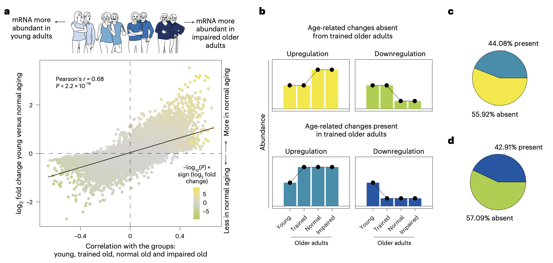
Figure 2: More than half of the molecular signature of muscle aging is absent in trained older adults. Age-related transcriptional changes correlated strongly with a muscle health gradient across the four groups. Between 45 and 62 percent of aging changes were absent in the trained group. The energy metabolism genes NDUFS1 and COX5A were depleted in normally active and impaired older adults but maintained youthful expression in the trained.
But the most important detail is not the fraction. It is which changes fell into the preventable category. The genes that training preserved at youthful levels were, overwhelmingly, the energy metabolism genes, the exact same cellular respiration signature that defined muscle aging in the first place. The strongest enrichment among the changes that training prevented was in genes for cellular respiration and energy production. NDUFS1, a core subunit of the first complex of the electron transport chain, was depleted in normally active and impaired older adults but sat at youthful levels in the trained. COX5A, a component of cytochrome c oxidase, showed the same pattern. Across mitochondrial respiratory complexes I through V, the trained older adults maintained expression levels resembling those of young adults, while the normally active and impaired groups showed the decline.
The significance of this alignment is hard to overstate. The single most prominent molecular change in muscle aging, the collapse of energy metabolism gene expression, turned out to be the single most preventable one. The thing that goes most wrong with aging muscle is the thing that structured training most effectively holds in place.
And it is worth returning to what the comparison group was, because it is what makes this finding remarkable rather than obvious. The trained older adults were not being compared to sedentary people. They were being compared to older adults who were walking just as much as young adults, matched step for step. General daily activity, the kind that fills a step counter, was not enough to preserve the energy metabolism signature. The normally active older adults were moving plenty and their mitochondrial gene expression declined anyway. What preserved it was structured, sustained, deliberate training, three hours a week of genuine exercise, maintained for years. That is a meaningfully different prescription from simply being active, and the molecular data draws the distinction sharply.
What Training Could Not Fix
If the story ended with more than half of muscle aging being preventable, it would be an inspiring result and an incomplete one. The more scientifically honest and ultimately more useful part of the study is the other half: the molecular changes that appeared in every older adult, trained or not, the changes that structured exercise did not touch.
These were the age-related changes that occurred across all older groups regardless of training status, and they were biologically distinct from the energy metabolism changes that training preserved. Two categories stood out.
The first was an upregulation of genes involved in synaptic transmission, the machinery by which nerve cells communicate with muscle. PCDH8, a protocadherin involved in cell-cell signaling at synapses, was elevated in all older adults. So was UNC13C, a gene involved in the release of neurotransmitters. The persistence of these changes across the trained group is notable because it points toward a dimension of muscle aging that is not fundamentally metabolic. The connection between nerve and muscle, the neuromuscular junction, deteriorates with age, and these transcriptional changes may reflect that deterioration. Whatever training does for the energy machinery of the muscle fiber, it did not reverse this signature of altered nerve-muscle communication.
The second was a downregulation of genes in the WNT signaling pathway, including DAAM2 and CTR9. WNT signaling governs a range of developmental and regenerative processes, including the behavior of the satellite cells that repair and maintain muscle tissue. Its decline across all older adults, trained included, suggests an age-related shift in the regenerative and developmental programs of muscle that structured exercise did not prevent.
The authors draw the crucial conceptual distinction directly. They divide age-related molecular changes into two categories: "preventable" changes, defined as those absent in trained older adults, and "unavoidable" changes, defined as those shared across all older groups regardless of how much they exercised. And they make an argument about what this division means for the field. The ambition of geroscience, they suggest, should be to develop therapeutics aimed at the unavoidable changes, the ones lifestyle cannot reach, while promoting exercise and lifestyle interventions to handle the preventable ones. This is a genuinely useful way to organize the problem. It tells you where behavior can do the work and where it cannot, and it tells drug developers where their efforts are actually needed rather than duplicating what a training program already accomplishes.
There is an important honesty in how the authors characterize the unavoidable changes. Unlike the preventable changes, which clustered cleanly around energy metabolism and cellular respiration, the unavoidable changes did not resolve into a single dominant pathway or a clean biological theme. They were scattered across synaptic, developmental, and regulatory processes without an obvious central mechanism. This matters because it means the therapeutic target is not yet obvious. The preventable half of muscle aging has a clear intervention, which is structured training. The unavoidable half does not yet have a clear molecular handle, and identifying one is precisely the work the authors argue the field should prioritize. Naming the problem is not the same as solving it, but knowing which half of aging is still waiting for a solution is a meaningful step toward finding one.
The Impaired Group: What Muscle Looks Like on the Way Down
If the trained older adults show what muscle aging looks like when it goes well, the physically impaired older adults show what it looks like when it goes badly. This group, the five older adults with Short Physical Performance Battery scores indicating meaningful functional decline, carried a molecular signature distinct from both the trained and the normally active, and it is worth examining because it illuminates the direction of travel toward frailty.
Two features defined the impaired group's muscle. The first was an elevation of immune and inflammatory pathway genes. Where the trained older adults showed enrichment in metabolic and lipid handling genes, the impaired group showed heightened expression of immune response pathways, a signature consistent with the chronic low-grade inflammation that accompanies frailty and that has been given the name inflammaging in the broader literature. The second was a depletion of mitochondrial genes, and specifically a striking loss of the genes encoding mitochondrial ribosomal proteins, including MRPS16, MRPL39, MRPL35, MRPL34, and MRPS18C.
This last detail is more mechanistically specific than it might first appear, and it is worth slowing down on. Cells contain two distinct populations of ribosomes, the molecular machines that manufacture proteins. Cytosolic ribosomes build the proteins of the general cell body. Mitochondrial ribosomes, built from a separate set of mitochondrial ribosomal proteins, are dedicated to manufacturing the specific proteins the mitochondria need to run the energy production machinery. In the impaired group, the mitochondrial ribosomal proteins were depleted while the cytosolic ribosomal proteins were not. The protein-manufacturing capacity of the mitochondria specifically was failing, while the general protein-manufacturing capacity of the cell was comparatively intact.
This is a meaningful distinction because it points to mitochondrial translation as a specific point of failure in declining muscle rather than a generalized collapse of cellular function. The muscle of the impaired older adults was not simply winding down across the board. It was losing, in a targeted way, the machinery required to build and maintain its energy-producing apparatus, compounding the same energy metabolism deficit that defined ordinary aging but going a step further into the specific breakdown of the systems that sustain mitochondrial function.
The contrast with the trained group runs in the opposite direction on every axis. Where the impaired group showed immune activation, the trained showed metabolic enrichment. Where the impaired group showed mitochondrial ribosomal depletion, the trained maintained mitochondrial gene expression near youthful levels. And in a structural confirmation of their endurance adaptation, the trained older adults showed a higher proportion of type I muscle fibers, the slow-twitch, fatigue-resistant, mitochondria-dense fibers that characterize aerobically trained muscle. The two groups were aging in opposite directions, and their muscle recorded the divergence in detail.
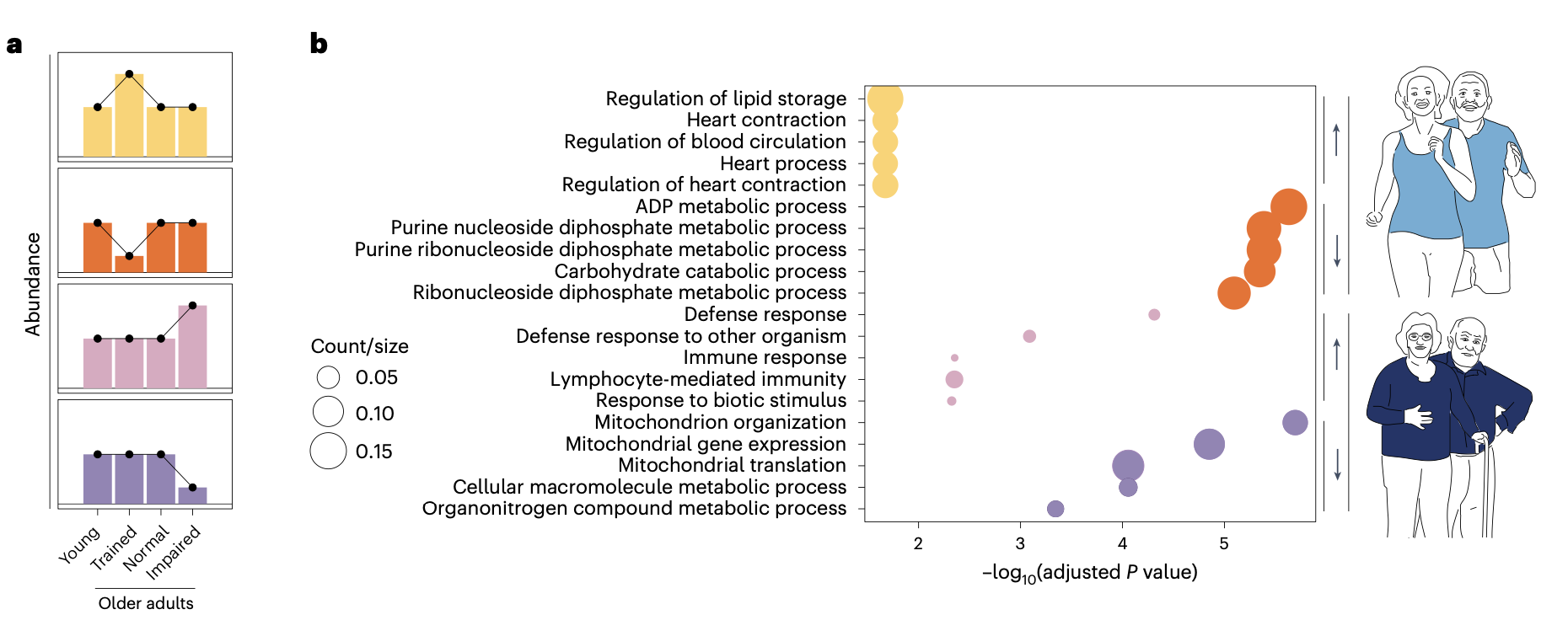
Figure 3: Trained and physically impaired muscle age in opposite directions. Trained older adults showed enrichment in lipid and metabolic genes, while impaired older adults showed elevated immune pathways and depleted mitochondrial genes. Mitochondrial ribosomal proteins including MRPS16, MRPL39, MRPL35, and MRPL34 were specifically depleted in the impaired group, while cytosolic ribosomal proteins were not, pointing to a targeted failure of mitochondrial protein synthesis.
The Counterintuitive Finding: Fitter Muscle Mounts a Bigger Stress Response to Exercise
Everything so far has concerned the resting state of the muscle, the baseline molecular profile of each group before exercise. But the study also captured something dynamic, and it produced the most surprising finding in the paper. When these participants actually exercised, the fitter they were, the larger the inflammatory and stress response their muscle mounted. And that larger response was associated with better physical function, not worse.
This runs against a common intuition. Inflammation is widely treated as something to be minimized, a marker of damage and dysfunction, and much of the longevity field is oriented toward suppressing it. The expectation might be that healthy, well-trained muscle would respond to a bout of exercise calmly, with minimal inflammatory disruption, while frail muscle would overreact. The study found the opposite.
After the one-hour cycling bout, all four groups activated a recognizable stress and immune response at the transcriptional level. Genes went up that anyone who studies inflammation would recognize: IL6, IL1B, and TNF, the classic inflammatory cytokines, along with SELE, an adhesion molecule involved in immune cell recruitment, and FOS, an immediate-early stress response gene. This activation was not unique to any one group. Every group, young and old, trained and impaired, mounted it. What differed was the magnitude, and the direction of that difference is the surprise.
The researchers measured how closely each older group's exercise response resembled the response of the young adults. The trained older adults were the closest match, with a correlation of 0.451. The normally active older adults were further off, at 0.394. And the physically impaired older adults were the most divergent, at 0.263, their muscle mounting the weakest and least youthful response to the same exercise stimulus. The capacity to respond vigorously to exercise tracked with fitness. The fitter the muscle, the more it resembled young muscle in how forcefully it reacted, and the more forcefully it reacted, the healthier the person tended to be.
The study went further and connected specific stress-response genes to specific measures of physical health. The team built a network correlating the exercise-induced change in each gene against physiological measurements taken at baseline. Several stress-response transcription factors emerged as central integrators. ATF2, a well-established target of the p38 MAPK stress signaling pathway and a regulator of mitochondrial biogenesis, showed exercise-induced changes that correlated with in vivo mitochondrial function. HEYL, involved in muscle regeneration and satellite cell activation, correlated with muscle strength. USP8, involved in protein quality control and cellular stress responses, correlated with mitochondrial function. In each case, a more robust exercise-induced stress response was associated with better underlying muscle health.
The interpretation the authors reach is that the stress response to exercise is not damage to be minimized. It is the mechanism of adaptation itself. Exercise works by stressing the muscle, and the muscle's ability to mount a vigorous stress response is what allows it to adapt and grow stronger. A muscle that responds forcefully to exercise is a muscle that is still capable of adapting to it. A muscle that responds weakly, as the impaired group's did, has lost some of that adaptive capacity. The stress response is not the cost of exercise. It is the point of it.
This leads the authors to a provocation that deserves to be stated in full, because it cuts against a significant current in longevity thinking. If the inflammatory and stress response to exercise is the mechanism through which exercise produces its benefits, then longevity interventions designed to suppress inflammation may carry a hidden cost. The authors name this concern directly, raising the possibility that interventions targeting immune suppression, such as the IL-11 inhibitors that have attracted attention as potential longevity drugs, or well-known anti-inflammatory agents taken chronically, might inadvertently blunt the very stress responses that drive functional resilience and adaptation in muscle. This is a hypothesis, not a demonstrated effect, and the study does not test any anti-inflammatory intervention directly. But the logic is coherent and the concern is legitimate. If you suppress the inflammatory signaling that exercise depends on to produce adaptation, you may weaken the adaptation itself. For an audience increasingly exposed to the idea that chronic inflammation is uniformly the enemy, this is an important and underappreciated complication. Some inflammation, in the right context and at the right time, is not damage. It is the signal that makes the body stronger.
The NAD+ Discovery: A New Regulator Hiding in the Data
Buried in the later analyses of this paper is a finding that has little to do with exercise and everything to do with one of the most discussed molecules in longevity science. Almost nobody reading the coverage of this study will know it is there, and it may prove to be one of the more consequential results in the paper. The researchers identified a previously unrecognized regulator of NAD+.
NAD+ is a molecule central to energy metabolism, required for the reactions that extract energy from fuel and for the activity of a family of enzymes involved in cellular maintenance and repair. Its levels decline with age across many tissues, including muscle, and this decline has made it one of the most heavily pursued targets in the longevity field. Supplements intended to raise NAD+, including precursors like nicotinamide mononucleotide, or NMN, have become widely used on the strength of that rationale. The question of how NAD+ is regulated in aging muscle is therefore of direct interest to a very large audience.
The team took advantage of the molecular depth of their dataset to ask which genes were associated with NAD+ levels. They correlated the expression of every transcript against the abundance of NAD+ and its related metabolites. The genes that correlated positively with NAD+ were, as expected, enriched for mitochondrial and respiratory chain processes, consistent with the well-established relationship between NAD+ and mitochondrial energy production. This was confirmation of known biology.
The genes that correlated negatively with NAD+ were the surprise. They were enriched for ubiquitin-mediated proteolysis, the cellular system responsible for tagging proteins for destruction and breaking them down through a structure called the proteasome. This was not an expected relationship. There was no established reason to think that the protein degradation machinery of the cell would be inversely related to NAD+ levels, and a correlation alone could not establish whether the relationship was causal or merely coincidental.
So the team tested it. They took the correlational hypothesis, that proteasome activity influences NAD+ levels, and subjected it to a direct experiment. They treated cultured muscle cells and liver cells with MG-132, a compound that inhibits the proteasome, and measured what happened to NAD+. If the negative correlation reflected a genuine causal relationship, then inhibiting the proteasome should raise NAD+.
It did. In C2C12 muscle cells, proteasome inhibition significantly increased NAD+ levels. The same effect appeared in AML12 liver cells, confirming it was not specific to muscle. And to calibrate the magnitude of the effect, the team compared it to treatment with NMN, the established NAD+ precursor supplement. Proteasome inhibition raised NAD+ to a degree comparable to NMN treatment. A manipulation that had nothing to do with supplying NAD+ building blocks, that simply slowed the destruction of proteins, produced an NAD+ increase on the same order as the leading precursor supplement.
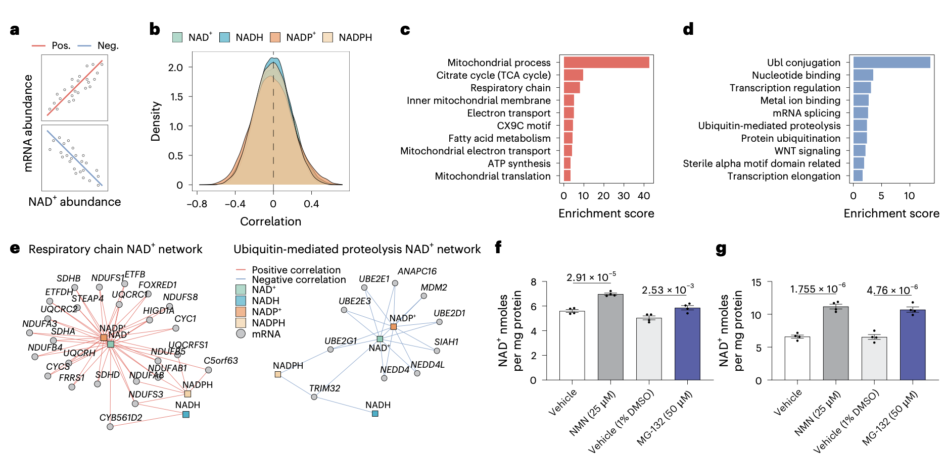
Figure 8: Proteasome inhibition raises NAD+ to a degree comparable to NMN. Genes negatively correlated with NAD+ were enriched for ubiquitin-mediated proteolysis. Testing this directly, the proteasome inhibitor MG-132 significantly increased NAD+ in both C2C12 muscle cells and AML12 liver cells, an effect comparable in magnitude to the established NAD+ precursor NMN, identifying protein degradation as a previously unrecognized regulator of NAD+ abundance.
The implication is that proteasome activity is a genuine regulator of NAD+ abundance, a lever on NAD+ levels that the field had not previously recognized. This does not mean that inhibiting the proteasome is a viable longevity strategy, and it should not be read that way. The proteasome performs essential functions, and broadly inhibiting it would carry serious consequences. But identifying it as a regulator of NAD+ opens a new line of inquiry into how NAD+ decline might be addressed, one that operates through protein turnover rather than through supplying more raw material. For a field that has focused heavily on precursor supplementation as the route to raising NAD+, the discovery that the degradation machinery of the cell also sets NAD+ levels is a genuinely new direction, and it emerged from a study that was ostensibly about exercise.
Limitations of the Study
The study's design is its greatest strength, but several limitations bound what it can establish.
The most important concerns the exercise itself. All participants exercised at 50 percent of their individual maximum capacity, which standardizes the relative effort but means the trained older adults performed more absolute work than the others, because their maximum was higher. Part of their stronger molecular response to exercise may therefore reflect the greater absolute workload rather than healthier aging per se. The two are difficult to fully separate.
The study is also cross-sectional, which carries a caution that applies to the headline finding. The trained older adults were not randomly assigned to years of training and compared against their own untrained baseline. They were people who had already trained for years. It is possible that they were biologically different before they ever began, that the people who become lifelong exercisers differ from those who do not in ways that would show up in muscle regardless of training. The study cannot prove that training caused the preserved molecular profile, only that the two are associated. The mechanistic coherence makes causation plausible, but a cross-sectional design cannot establish it.
Several other limitations are worth noting briefly. The impaired group was small, only five participants, though recruiting and biopsying pre-frail older adults is genuinely difficult and their inclusion is a strength despite the number. The transcriptomics was performed on bulk tissue rather than at single-cell resolution, which limits the ability to attribute changes to specific cell types, a limitation compounded by the trained group's higher proportion of type I fibers. The study was not powered to detect sex differences, though the authors note the respiration decline may be more pronounced in women. And the cohort was Dutch, drawn from a population with unusually high baseline activity levels, so the findings warrant validation in more sedentary populations before being generalized.
None of these undermines the central results. But they define the appropriate confidence: this is a rigorous and unusually well-designed cross-sectional study that identifies strong associations and a plausible causal story, not a randomized trial that proves training rewrites the aging trajectory.
What This Means
Strip away the molecular detail and this study delivers three messages that matter for how a person thinks about aging, exercise, and the interventions marketed to address both.
The first is that the most prominent feature of muscle aging is substantially preventable, but the prescription is more specific than most people assume. The collapse of energy metabolism gene expression, the single largest molecular signature of aging muscle, was largely absent in the trained older adults. But it was fully present in the normally active ones, and those normally active older adults were walking as much as the young adults. General daily movement, the kind that fills a step counter and satisfies most public health guidelines, was not enough to preserve the mitochondrial signature. What preserved it was structured, sustained training, three genuine sessions a week, held for years. This is a meaningful distinction. Being generally active and being trained are not the same thing at the molecular level, and this study suggests the difference between them is much of what separates muscle that ages well from muscle that does not.
The second is that there is a floor, and knowing where it sits is valuable. Roughly half of the molecular signature of muscle aging persisted regardless of training. These unavoidable changes, in synaptic and developmental and regulatory genes, are where lifestyle runs out of road. No amount of exercise in this study touched them. That is not a discouraging finding, it is a clarifying one. It tells the person doing the training that they are addressing the preventable half as effectively as anything currently available, and it tells the geroscience field that the unavoidable half is where drugs will have to do the work, because behavior cannot. The honest complication is that this unavoidable half does not yet resolve into a clean target, which means the work of finding one is still ahead.
The third message is the most immediately provocative, and it should give pause to anyone drawn to the idea that inflammation is uniformly the enemy. The inflammatory and stress response that muscle mounts in response to exercise appears to be the mechanism of adaptation, not a side effect to be minimized. The fitter the muscle, the larger that response, and the larger the response, the better the physical function. This raises a genuine concern about the growing interest in chronic anti-inflammatory interventions for longevity, including IL-11 inhibitors and the routine use of anti-inflammatory agents. If the stress response to exercise is what makes exercise work, then suppressing it systemically may quietly undercut one of the most powerful longevity interventions available. This does not mean inflammation is good, or that anti-inflammatory strategies have no place. It means the timing and context matter enormously, and that blunting the acute, exercise-induced stress response may carry a cost that a simple more-inflammation-is-worse model does not capture.
Taken together, these findings support a view of exercise not as a general wellness recommendation but as a molecularly specific intervention that does specific things, reaches specific targets, and leaves others untouched. It is more powerful than a step count and more precise than a platitude, and this study is among the clearest accounts yet of exactly what it does inside the tissue.
Conclusion: A Map With Two Territories
For decades, the conversation about exercise and aging has been conducted largely in generalities. Exercise is good for you. Movement preserves function. Stay active as you age. All of this is true, and all of it is too vague to tell you what exercise actually does or where its limits lie. This study replaces the generalities with a map.
The map has two territories. One is the preventable half of muscle aging, dominated by the decline of energy metabolism, and the study's central finding is that this territory is more changeable than almost anyone expected. More than half of the molecular signature of aging muscle was absent in people who had trained consistently for years, and the part that was absent was precisely the part that matters most for how muscle produces energy and sustains function. This is the territory where behavior wins, where structured training does work that no drug currently matches, and where the evidence says the ceiling on what a person can preserve is higher than the resigned narrative of inevitable decline would suggest.
The other territory is the unavoidable half, the changes that appeared in everyone regardless of how much they trained. This is the territory where lifestyle runs out and where the future of geroscience will have to operate. The study does not solve this half. It does something arguably more useful at this stage, which is to draw the border clearly, to say with molecular precision where exercise stops being able to help and where new interventions will be needed. Knowing where that border sits is what allows the field to aim its efforts where they are actually required rather than duplicating what a training program already achieves.
And running through the whole map is the reminder that the stress response to exercise, the acute inflammatory signal that a hard bout of activity provokes, is not damage to be suppressed but the mechanism through which the benefit is delivered. The fittest muscle mounted the largest response, and that should make anyone reaching for a chronic anti-inflammatory in the name of longevity pause to consider what they might be turning down along with the inflammation they mean to address.
What the study ultimately provides is a molecular atlas of fitness-dependent aging, a resource that future work will mine for years, and a clearer answer than we have had to a question people have been asking for as long as they have been growing older: how much of this is up to me? The answer is more than you might think, and not all of it, and the value of this study is that it tells you, with unusual precision, which is which.
- Janssens, G.E., Trętowicz, M.M., Grevendonk, L. et al. Delayed molecular aging, preservation of energy metabolism and enhanced exercise response in exercise-trained human muscle. Nat Aging 6, 1482–1500 (2026). https://doi.org/10.1038/s43587-026-01150-x
Related studies